| Team Members: | Mary Aronne1, Samantha Clapp2, Soohwan Jung3, Abigail Kramer4, and William Wang5 |
| Graduate Assistant: | Janita Patwardhan1 |
| Faculty Mentor: | Bradford E. Peercy1 |
| Client: | Arthur Sherman6 |
1Department of Mathematics and Statistics, University of Maryland, Baltimore County,
2Department of Mathematics, Georgia College and State University,
3Department of Mathematics, Edmonds Community College,
4Department of Mathematical Sciences Kent State University,
5Department of Mathematics, Vanderbilt University,
6Laboratory of Biological Modeling, National Institutes of Health
About the Team
Team 4 consists of five undergraduate students participating in the summer 2016 Interdisciplinary High Performance Computing REU at UMBC. Mary Aronne is a junior at UMBC and is majoring in mathematics with a minor in environmental science. Samantha Clapp is a senior at Georgia College and State University and is studying mathematics with a French minor. Soohwan Jung is a junior from Edmonds Community College and is transferring to UCLA. He studies applied mathematics. Abigail Kramer is a senior at Kent State University and studies applied math with a computer science minor. William Wang is a sophomore at Vanderbilt University and is a mathematics major. Team 4 worked with graduate assistant Janita Patwardhan and faculty mentor Bradford Peercy in the Department of Mathematics and Statistics at UMBC. The client, Arthur Sherman, is from the Laboratory of Biological Modeling at the National Institutes of Health.
Problem
Diabetes is a disease characterized by an excessive level of glucose in the bloodstream, which may be a result of improper insulin secretion. Insulin is secreted in a bursting behavior of pancreatic β-cells in the islets of Langerhans, which is affected by oscillations of cytosolic calcium concentration. We used the Dual Oscillator model to explore the role of calcium in calcium oscillation independent (CaI) versus calcium oscillation dependent (CaD) modes as well as the synchronization of metabolic oscillations in electrically coupled cells.
Dual Oscillator Model
The Dual Oscillator Model (DOM) provided by Dr. Sherman consists of seven differential equations which simulate a single pancreatic β-cell. In order to simulate a greater number of β-cells to represent an islet, a diagonal coupling matrix is utilized, which connects the voltages and calcium concentrations between a cell and its neighbors.

Synchronization
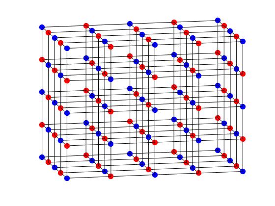
We simulated heterogeneous islet models of different bursting cells to evaluate the synchronization of the cells in each islet model. We used an islet cube model where two JGK (flux of glucokinase) values are equally distributed. We took JGK values from the CaD and CaI regions, and categorized each JGK pair as CaI, Mixed, and CaD. Mixed represents where the one JGK value of the islet model is CaI and the other is CaD.
We observed from our simulations of the CaD modes that increasing voltage coupling increases synchronization.
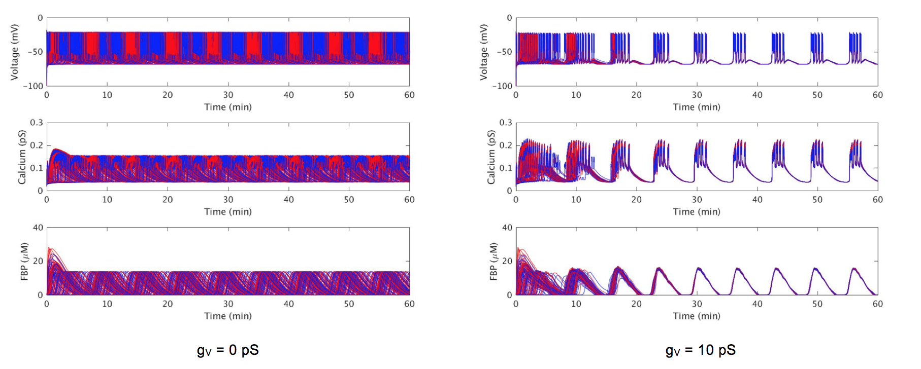
is 0.18 μM · ms-1 (blue) or is 0.2 μM · ms-1 (red)
We obtained synchronization indices for the above islet cube model where we altered the voltage and calcium coupling strengths.
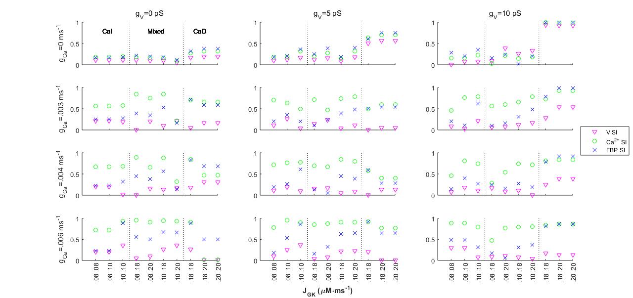
The above chart consists of twelve scatter plots with each one representing a different voltage and calcium coupling combination. The x-axis shows the combinations of JGK values while the y-axis represents the synchronization index (SI) values. To represent the SI values of voltage, calcium and FBP per simulation, each point is given a certain shape and color, voltage synchronization being a pink triangle, calcium synchronization a green circle, and FBP synchronization a blue X. Synchronization index of 1 represents complete synchronization.
Conclusions
Coupling complex cells together has interesting dynamic effects. When in CaI modes, increasing calcium coupling with no voltage coupling increases synchronization. In CaD modes, increased voltage coupling with no calcium coupling increases synchronization. However, in CaD modes, voltage coupling with high calcium coupling causes desynchronization in voltage.
Links
Mary Aronne, Samantha Clapp, Soohwan Jung, Abigail Kramer, William Wang, Janita Patwardhan, Bradford E. Peercy, and Arthur Sherman. The Interaction of Calcium and Metabolic Oscillations in Pancreatic Beta Cells. Technical Report HPCF-2016-14, UMBC High Performance Computing Facility, University of Maryland, Baltimore County, 2016. (HPCF machines used: maya.). Reprint in HPCF publications list
Poster presented at the Summer Undergraduate Research Fest (SURF)
Click here to view Team 1’s project
Click here to view Team 2’s project
Click here to view Team 3’s project
Click here to view Team 5’s project
Click here to view Team 6’s project
Click here to view Team 7’s project